New tests available in S’pore to help assess adults for ADHD
SINGAPORE – Adults who suspect they have attention deficit hyperactivity disorder (ADHD) can now undergo a new series of tests designed to provide a more


SINGAPORE – Adults who suspect they have attention deficit hyperactivity disorder (ADHD) can now undergo a new series of tests designed to provide a more

Keynotes from leading experts in the field: David Mehler, MD, PhD (Applied Computational Neuroscience (ACN) lab at the Uniklinik RWTH Aachen and Lucas R. Trambaiolli,
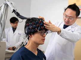
the fNIRS community recognised the need for a state-of-the-art fNIRS data preprocessing and analysis software. Satori is our answer to these needs developed by NIRx
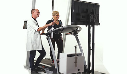
Cerebral palsy (CP) is a non-progressive neurologic condition that causes gait limitations, spasticity, and impaired balance and coordination. Robotic-assisted gait training (RAGT) has become a

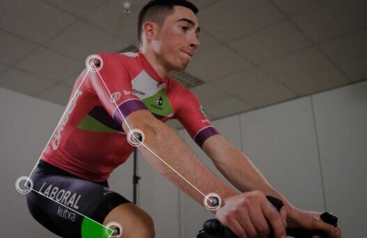
An improvement in ecological validity is one of the significant challenges for 21st-century neuroscience. At the same time, the study of neurocognitive processes in real-life
Imagine a 4 year old doing a MRI exam without the need for anesthesia, impossible right? Watch this interview featuring radiographer and MR technician Bac
Today marks the Longest Day, an event that marks the Summer solstice that encaptures family, friends, and patients with the goal to fight the darkness